Apr 212014
 

Original story by Warren Barnsley, Sydney Morning Herald

Budding young filmmakers are being encouraged to shoot video evidence of marine debris affecting the Great Barrier Reef in a bid to raise awareness of the issue.

Great Barrier Reef

The Gladstone Local Marine Advisory Committee is calling on eight to 18-year old documentary producers to put together short films highlighting the problem of marine debris.

“We want young people to use their creativity to tell a compelling story about marine debris in a video no longer than two minutes,” said Gladstone LMAC Chair Blue Thomson.

“It can be an interview, documentary-style, a music video, a fictional story or animated. It’s entirely up to the creator,” he said.

Researchers say it’s a major issue for the world heritage-listed ecosystem, not only because of the negative impacts to the reef’s aesthetic qualities and hazard to ocean users.

LMAC member and Central Queensland University Research Fellow Dr Scott Wilson claims plastics are a top five pollutant causing harm to the marine environment and animals.

“In a recent study, 22 per cent of shearwater chicks were found to have plastics in their stomachs.

“Plastic bags, bottles, ropes and nets trap, choke, starve and drown many marine animals and seabirds around the world every year.”

The issue could be better dealt with if people were more responsible with their litter, including plastics, rubbers, metal, wood and glass, said Dr Wilson.

Participants will go in the running to win an iPad or GoPro Hero 3, with entries closing on May 30.

Winners will be announced on June 16.

Apr 192014
 

Media release from  Max-Planck-Gesellschaft

Newly discovered types of neurons in the animals’ brain help to compensate for self-motion

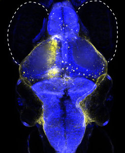
Newly discovered neuron type (yellow) helps zebrafish to coordinate its eye and swimming movements. Photo: © Max Planck Institute of Neurobiology/Kubo

Newly discovered neuron type (yellow) helps zebrafish to coordinate its eye and swimming movements. Photo: © Max Planck Institute of Neurobiology/Kubo

Our eyes not only enable us to recognise objects; they also provide us with a continuous stream of information about our own movements. Whether we run, turn around, fall or sit still in a car – the world glides by us and leaves a characteristic motion trace on our retinas. Seemingly without effort, our brain calculates self-motion from this “optic flow”. This way, we can maintain a stable position and a steady gaze during our own movements. Together with biologists from the University of Freiburg, scientists from the Max Planck Institute of Neurobiology in Martinsried near Munich have now discovered an array of new types of neurons, which help the brain of zebrafish to perceive, and compensate for, self-motion.

When we jog through a forest, the image of the trees appears to move backwards across our retina. This occurs for both eyes in the same direction. If, however, we turn about our own axis, the trees appear to rotate around us. For one eye, this rotation goes from the outside in, and for the other one it goes from the inside out. Our brain processes such large-scale movements in the visual environment, the “optic flow”, so that when jogging, for example, we can estimate our speed correctly and do not constantly stumble.

The human brain is, of course, not unique in being able to perceive optic flow. Fish that live in rivers and streams use this capability, for example, to prevent themselves from drifting in the current. Based on the optic flow, the fish corrects its passive drifting through its own swimming. How and where the fish brain carries out these calculations was not previously known.

“We wanted to know how the compensatory movements are triggered and by which neurons,” explains Herwig Baier. Together with his department at the Max Planck Institute of Neurobiology, he searches for and describes the neural networks in the brains of zebrafish larvae that control certain types of behaviour. This is no easy task, as, despite its minuscule size, the brain of a 5-mm-long fish larva consists of several hundred thousand neurons. One advantage, however, is that the brain of the fish larva is almost completely transparent. Neurons can thus be observed directly under the microscope without requiring any surgical dissection.

For their experiments, the scientists placed the fish larvae in circular containers, where they saw black-and-white stripes that moved around them. The animals demonstrated different reactions depending on the movement pattern presented. When the stripes moved from back to front for both eyes, the fish swam straight ahead or tried to turn around. However, when the stripes moved around the fish in a clockwise or counter-clockwise direction, the two eyes followed the perceived direction of rotation. The compensatory movements of the entire body (optomotor behaviour) or of the eyes alone (optokinetic behaviour) should make the motion signal on the retina as small as possible – and keep the fish stable in place.

The neurobiologists wanted to identify the neurons while the brain was processing self-motion and initiating optomotor or optokinetic movements. “It was like looking for a needle in a haystack,” explains Fumi Kubo, first author of the study. “This would have been completely inconceivable just a few years ago.” For her study, Fumi Kubo, who worked in collaboration with Aristides Arrenberg and Wolfgang Driever from the Institute of Biology I at the University of Freiburg and scientists from the Freiburg Cluster of Excellence BIOSS Centre for Biological Signalling Studies, used a new scientific method: the imaging of the entire brain. Thanks to the latest fluorescent dyes and sophisticated genetic techniques, it has recently become possible to visualise the outlines of all neurons in a fish brain. The special feature of this technique, however, is that the dyes change colour when a neuron becomes active.

During the experiment, the heads of the fish with the labelled nervous system were embedded in a gel. The moving striped patterns on the walls of the container gave the animals the impression of self-motion, similar to the sensation triggered in an IMAX cinema. Depending on whether the stripes drifted forward or rotated, the fish followed the patterns with their eyes or beat their tails. Using a two-photon microscope, the scientists were able to observe which neurons reacted to the direction of the moving stripes.

Four direction-selective cell types had previously been identified in the retina. For a long time, scientists had predicted that these cells somehow carry information about optic flow to downstream neurons in the visual brain, which in turn transmitted the commands to the motor centers that control eye and body movements. The neurobiologists have now succeeded in demonstrating the existence of such comparatively simple neuronal connections. They also discovered seven previously unknown cell types responsible for more complex responses to the inputs from both eyes. For example, one type of cell becomes active when both eyes perceive a forward movement but not a clockwise rotation, which would evoke a turn to the right. This finding is remarkable as in both cases, the left eye should detect a movement from the outside in. “So, not only did we find new cell types, we also discovered a possible explanation as to why the fish’s brain distinguishes between translational (that is, forward or backward) and rotational (that is, clockwise or counterclockwise) movements,” explains Fumi Kubo.

Once the fish were placed back, swimming freely in their tank, the scientists produced a wiring diagram of the cells based on the recorded tasks for the new neuron types and their locations in the brain. Their findings help to provide a better understanding of the processing of movements in the vertebrate brain. However, Fumi Kubo is already thinking about the next stage in the research: “The next challenge will be to prove the proposed connections in the brain.”

Apr 192014
 

Original story by Celeste Biever and Lisa Grossman, New Scientist

The pitch has dropped – again. This time, the glimpse of a falling blob of tar, also called pitch, represents the first result for the world’s longest-running experiment.

Sadly however, the glimpse comes too late for a former custodian, who watched over the experiment for more than half a century and died a year ago.

Up-and-running since 1930, the experiment is based at the University of Queensland in Australia and seeks to capture blobs of pitch as they drip down, agonisingly slowly, from their parent bulk.

It was pipped to the post last year when a similar experiment, set up in 1944 at Trinity College Dublin in Ireland, captured the first ever video footage of a blob of pitch droppingMovie Camera.

In that instance, the blob separated from its parent bulk. By contrast, the Australian team filmed the collision between the ninth blob ever to fall and the eighth blob, which was sitting at the bottom of their beaker – but the ninth blob is still attached to the pitch above it.

Still, the Australian result is important because the experiment has a better set-up, says Stefan Hutzler, a member of the Trinity College Dublin team who used those results to calculate the pitch’s viscosity. “Theirs is in a glass container; they measure the temperature, measure the humidity as well,” he says. “Ours, we don’t really call it an experiment. It was really just sitting there on a shelf, going back to the 1940s.”

Near miss

The fact that both experiments dropped within a year of each other is “just pure luck”, says Hutzler. Hot summer weather in Ireland last year may have influenced the timing.

The Queensland experiment already features in the Guinness World Recordsand won an IgNobel prize in 2005. It was set up by physicist Thomas Parnell to illustrate that although pitch appears solid, shattering when hit with a hammer at room temperature, it is actually a very viscous liquid.

The eventual result follows several near misses, according to the University of Queensland. John Mainstone, who oversaw the experiment for more than 50 years until his death last August, missed observing the drops fall three times – by a day in 1977, by just five minutes in 1988 and, perhaps most annoying, in 2000, when the webcam that was recording it was hit by a 20-minute power outage.

“It’s a pity of course that the person in charge died about a year ago, so he never saw the drop,” Hutzler says. “He would have enjoyed that.”

Honey flow

The eighth and ninth drops each took about 13 years to fall, says current custodian Andrew White. By contrast, the seven drops that fell between 1930 and 1988 did so faster – at an average rate of one drop every eight years.

The next step is to see how long it takes the ninth drop to separate from the pitch above it: “It may tip over quickly or it might slow right down and take years to break away,” says White.

You can keep an eye on the ninth drop’s movements via a live web stream. The University of Queensland says it will work out who was watching when the pitch dropped and record their names for posterity.

The drop experiments show that the physics of a drop forming in a viscous material is still not well understood, Hutzler says – although he doesn’t think watching pitch for decades is necessarily the best way to study it. Using honey or some other less viscous fluid would give you better statistics.

“I think these experiments capture the imagination just because they go on for such a long time,” he says. The video of the drop in Dublin quickly went viral on YouTube. “Ironically, you have a very slow event happening, but the news spreads very quickly.”

Mar 302014
 

A video by Jose Lachat that won the “Prix de ‘l Insolite” (the prize of the unusual) at the Festival Mondial de l’Image Sous-Marine in Antibes, France.

The Hairy Frogfish (Antennarius striatus) swallows fishes taller than himself.

Video by Jose Lachat.
Festival Modial de l’Image Sous-Marine Antibes
“Prix de ‘l Insolite”

Video: Jose Lachat
Musik: Maia Wackernagel
www.wackernagel.ch
Schnitt: Claudius Buser

Mar 192014
 

Original story by Ben Chenoweth, the Wollondilly Advertiser

ANDREW Bodlovich’s kitchen may not “rule” but his barramundi and herb farm operation is certainly proving successful.
Herb farmer's insights: Andrew Bodlovich at his Cobbitty herb and barramundi farm. Photo: Jonathan Ng

Aquaponic herb farmer’s insights: Andrew Bodlovich at his Cobbitty herb and barramundi farm. Photo: Jonathan Ng

The 50-year-old Cobbitty farmer was chosen — as one of 34 farmers who supply Coles supermarkets — to have a brief guest appearance on Channel Seven’s reality show My Kitchen Rules.

Mr Bodlovich said his inclusion in the show, due to air on March 24, was to give the contestants and viewers an insight of where the produce originated.

“I think it’s really important for people to see beyond the cooking challenge,” he said.

“It’s a cooking show but they also want to showcase where the food comes from.”

It will be the second time the local farmer has graced Australian television screens.

His first appearance was on the ABC’s New Inventors program, where he and a fellow inventor demonstrated their combined herb farming and barramundi technique.

Mr Bodlovich said the system was designed so both fauna and fish benefited from the other.

“It’s all inside a high-tech glasshouse,” he said. “The vegetables are on a conveyor belt and below are the fish in tanks. The fish produce the nutrients and it turns into plant food. The fish feed the plants [which] keep the water clean.”

Mar 162014
 

Original story from International Science Times

MIT unveiled a robotic fish this week, a soft, silicone machine that can move autonomously through water. In the new journal Soft Robotics, MIT researchers describe the robot’s ability to execute escape manoeuvres like a real fish, turning 100 degrees in as many milliseconds, with the help of a carbon dioxide canister and a heap of machinery in the “brains” of the fish. The robot may eventually be used to swim in real fish colonies in order to gather data on their behaviour.

“Because of their body’s capability to bend and twist, these robots are capable of very compliant motion, and they are also capable of very rapid, agile maneuvers, which pushes the envelope on what machines can do today,” says MIT researcher Daniela Rus in the video below.

The inside of the fish is made up of control units in the head, a carbon dioxide canister in the head and abdomen and tubes which go from the CO2 canister to the tail. Changes in the level of CO2 determine how fast the fish moves, and the amount the tubes inflate changes the fish’s angle. The soft robot can be directed by an operator via a wireless receiver in the fish’s head. Silicone rubber (waterproof, of course) covers the outside of the fish.

Soft robots can offer several advantages over so-called hard robots (think of pretty much every other robot you’ve ever seen). Most robotsprioritize avoiding collisions–they don’t want to be damaged or fall over–which means that they may take an inefficient path to get where they’re going. But soft robots can withstand collision, and may even benefit from knocking into something.

“In some cases, it is actually advantageous for these robots to bump into the environment, because they can use these points of contact as means of getting to the destination faster,” says Rus. “The fact that the body deforms continuously gives these machines an infinite range of configurations, and this is not achievable with machines that are hinged.”

Last year, Rus and her colleagues showed off another incredible robotics project made up of colored blocks that spin and build themselves into modular machines. These building-block robots have to be seen to truly understood, so check them out.


Mar 102014
 

Original story by Richard Ingham/AAP at the Sydney Morning Herald

The space rock that smashed into Earth 65 million years ago, famously wiping out the dinosaurs, unleashed acid rain that turned the ocean surface into a witches’ brew, researchers said on Sunday.
An artist's impression of the recently identified Torvosaurus gurneyi dinosaur. Photo: Reuters

An artist’s impression of the recently identified Torvosaurus gurneyi dinosaur. Photo: Reuters

Delving into the riddle of Earth’s last mass extinction, Japanese scientists said the impact instantly vapourised sulphur-rich rock, creating a vast cloud of sulphur trioxide (SO3) gas.

This mixed with water vapour to create sulphuric acid rain, which would have fallen to the planet’s surface within days, acidifying the surface levels of the ocean and killing life therein.

Those species that were able to survive beneath this lethal layer eventually inherited the seas, according to the study which did not delve into the effects on land animals.

“Concentrated sulphuric acid rains and intense ocean acidification by SO3-rich impact vapours resulted in severe damage to the global ecosystem and were probably responsible for the extinction of many species,” the study said.

The great smashup is known as the Cretaceous-Tertiary extinction.

It occurred when an object, believed to be an asteroid some 10 kilometres wide, whacked into the Yucatan peninsula in modern-day Mexico.

It left a crater 180 kilometres wide, ignited a firestorm and kicked up a storm of dust that was driven around the world on high winds, according to the mainstream scenario.

Between 60 and 80 per cent of species on Earth were wiped out, according to fossil surveys.

Large species suffered especially: dinosaurs which had roamed the land for some 165 million years, were replaced as the terrestrial kings by mammals.

Extinction riddle

Much speculation has been devoted to precisely how the mass die-out happened.

A common theory is that a “nuclear winter” occurred – the dust pall prevented sunlight reaching the surface, causing vegetation to shrivel and die, and dooming the species that depended on them.

Another, fiercely debated, idea adds acid rain to the mix.

Critics say the collision was far likelier to have released sulphur dioxide (SO2) than SO3, the culprit chemical in acid rain. And, they argue, it would have lingered in the stratosphere rather than fallen back to Earth.

Seeking answers, a team led by Sohsuke Ohno of the Planetary Exploration Research Centre in Chiba set up a special lab rig to replicate — on a tiny scale —what happened that fateful day.

They used a laser beam to vapourise a strand of plastic, which released a high-speed blast of plasma and caused a tiny piece of foil, made of the heavy metal tantalum, to smash into a sample of rock.

The heavy foil fragment replicated on a miniscule scale the mass of the asteroid, while the rock was of a similar makeup as the surface where the asteroid struck.

The team caused collisions ranging from 13 to 25 km per second (47,000-90,000 km per hour), and analysed the gas that was released.

The research, reported in the journal Nature Geoscience, showed that SO3 was by far the dominant molecule, not SO2.

The team also carried out a computer simulation of larger silicate particles that would have been ejected by the impact, and found they too played a part.

The articles rapidly bound with the poisonous vapour to become sulphur acid “aerosols” that fell to the surface.

Heavily acidic waters would explain the overwhelming extinction among surface species of plankton called foraminifera.

Foraminifera are single-celled creatures protected by a calcium carbonate shell, which dissolves in acidic water.

The “acid rain” scenario also helps explain other extinction riddles, including why there was a surge in the number of ferns species after the impact. Ferns love acidic, water-logged conditions such as those described in the study.

Mar 082014
 

Original story by John Ross, The Australian

AN expert on Sydney Harbour’s marine life has taken out a new award from the Australian Academy of Science.

University of NSW marine ecology professor Emma Johnston, inaugural winner of the Australian Academy of Science's Nancy Millis Medal for Women in Science. Photo: Supplied

University of NSW marine ecology professor Emma Johnston, inaugural winner of the Australian Academy of Science’s Nancy Millis Medal for Women in Science. Photo: Supplied

The academy has marked International Women’s Day today by presenting its inaugural Nancy Millis Medal for Women in Science to University of NSW marine ecologist Emma Johnston.

The award is for early-and mid-career women scientists who have established independent research programs and demonstrated exceptional leadership in any branch of the natural sciences.

Professor Johnston is a faculty member at UNSW’s School of Biological, Earth and Environmental Sciences. She also heads the Sydney Harbour Research Program at the Sydney Institute of Marine Science, a collaboration of universities and government agencies.

The five-year project aims to help inform the management of the harbour’s natural and economic resources. Professor Johnston said the harbour was one of the most biologically rich in the world.

“Below the surface we find extensive kelp forests, sweeping seagrass meadows, rocky reefs and vibrant sponge gardens all teeming with life. (But) humans have used oceans for waste disposal for generations because they have little emotional attachment to what’s under the water.

“We need to get political will and resources going to clean it up.”

Professor Johnston’s research focus is the effects of pollutants on estuarine life, taking both an ecological and ecotoxicological perspective and “using field experimentation wherever possible”, her web page says.

Her research has taken her from the tropical waters of the Great Barrier Reef to Antarctica, where she has studied the impact of climate change on ecosystems on the polar seabed.

UNSW deputy vice-chancellor (research) Les Field, who is also the academy’s secretary for science policy, said Professor Johnston was a deserving recipient.

“Emma is a research powerhouse in marine science and an academic leader at UNSW as well as being an excellent role model to younger scientists, both here and across Australia.”

Mar 062014
 

Press release from  The University of Chicago Medical Center

Alternative routes to the same evolutionary destination: repeated origins of new fins in teleost fishes.

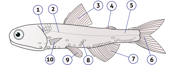
Anatomy of a Teleost fish - Lampanyctodes hectoris (Hector's lanternfish). (1) - operculum (gill cover), (2) - lateral line, (3) - dorsal fin, (4) - adipose fin, (5) - caudal peduncle, (6) - caudal fin, (7) - anal fin, (8) - photophores, (9) - pelvic fins (paired), (10) - pectoral fins (paired). Image: Lukas3/Wikimedia Commons

Anatomy of a Teleost fish – Lampanyctodes hectoris (Hector’s lanternfish). (1) – operculum (gill cover), (2) – lateral line, (3) – dorsal fin, (4) – adipose fin, (5) – caudal peduncle, (6) – caudal fin, (7) – anal fin, (8) – photophores, (9) – pelvic fins (paired), (10) – pectoral fins (paired). Image: Lukas3/Wikimedia Commons

Though present in more than 6,000 living species of fish, the adipose fin, a small appendage that lies between the dorsal fin and tail, has no clear function and is thought to be vestigial. However, a new study analyzing their origins finds that these fins arose repeatedly and independently in multiple species. In addition, adipose fins appear to have repeatedly and independently evolved a skeleton, offering a glimpse into how new tissue types and structural complexity evolve in vertebrate appendages.

Adipose fins therefore represent a prime example of convergent evolution and new model for exploring the evolution of vertebrate limbs and appendages, report scientists from the University of Chicago in theProceedings of the Royal Society B on March 5.

“Vertebrates in general have conserved body plans, and new appendages, whether fins or limbs, evolve rarely,” said senior author Michael Coates, PhD, chair of the Committee on Evolutionary Biology at the University of Chicago. “Here, we have a natural experiment re-run repeatedly, providing a superb new system in which to explore novelty and change.”

Usually small and structurally simple, adipose fins tend to get attention only when they are clipped from farm-raised trout and salmon as a tag. Despite their presence in thousands of fish species, they have been dismissed as a remnant of a once-functional fin. This assumption puzzled Coates and his co-authors, as they saw no evidence of deterioration in adipose fin structure or function in the fossil record.

To study the evolutionary origins of this fin, Coates and lead author Thomas Stewart, graduate student in organismal biology and anatomy at the University of Chicago, turned to a technique known as ancestral-state reconstruction. With co-author W. Leo Smith, PhD, from the Biodiversity Institute at the University of Kansas, they created an evolutionary tree describing the relationships between fish with and without adipose fins, using genetic information from more than 200 ray-finned fish and fossil data from known time points. They then used statistical models to predict when and in what species the adipose fin might have first evolved.

They found that adipose fins originated multiple times, independently, in catfish and other groups of ray-finned fishes — a striking example of convergent evolution over a vast range of species.

“It’s pretty incredible that a structure which is incredibly common could be so misunderstood,” Stewart said. “Our finding, that adipose fins have evolved repeatedly, shows that this structure, long assumed to be more-or-less useless, might be very important to some fishes. It’s exciting because it opens up new questions.”

More than 600 species of fish were studied in the course of this research, including many from the collections of the Field Museum in Chicago. This analysis revealed that a number of complex skeletal structures, including spines, plates, fin rays and cartilage discs, evolved independently in the adipose fins of different species. And while studies of the fossil record have suggested that new fins originate in a predictable and repeated manner, adipose fins demonstrate multiple routes to building new appendages.

“These results challenge what was generally thought for how new fins and limbs evolve, and shed new light on ways to explore the full range of vertebrate limb and fin diversity,” Stewart notes.

The study, “The origins of adipose fins: an analysis of homoplasy and the serial homology of vertebrate appendages,” was supported by the National Science Foundation and the University of Chicago Division of Biological Sciences.